Cone beam multi-angles radiography
*190�°orbital rotation and continuous pulse exposure realize Maximum density projection.
*Multi-angle dynamic continuous images on same part can be acquired.
*Replay the whole rotation dynamic images,volume rendering technique(VRT).
*Multi-angles observation during prepperative,intraoperative and postoperative,ensure accurate and efficient diagnosis and treatment.
*Choose appropriate sequence frames according to clinical need.both image quality and radiation protection can be assured.
Application
*Widely use in Orthopedics,general surgery,orthopedic surgery,urology,spinal surgery, abdominal surgery,gastroenterology department,gynecology and operating room etc.
*Bone tissue biopsy,spine pedicle screw implantation,long tubal bone marrow nail fixation and hand,foot fracture surgery with screw fixation.
Precise imaging and Perfect visualization
*Digital High frequency X-ray generator with micro-focus improve resolution and sharpness.
*Intraoperative 3D imaging enables intraoperative revisions and evaluation and replaces postoperative CT control.
*High frequency technology generator assures the best image in every operating condition along with reduced patient dose and extended x-ray tube lifetime.
X ray tubedual-focus fixed anode:0.6mm/1.5mm
Italy imd technology
Image intensifierthree fields of vision 9, 6, 4.5 french thales brand
Ccd cameraccd camera, japanese watt brand, 360rotation
Image systemrecursive noise reduction
Frame freezing
Multi frame image storage
Image conversing and rotating
Screen display with image comparison
Mechanical motion specificationsverticaltravel:0-400mm horizontaltravel:0~200mm rotation about horizontal axis:180
C-arm dimensiondistance of focal spot to image generator(sid):1000mm
Depth in arm 650mm
Mobile stand :1800mm*800mm*1850mm
Image system: 750mm*530mm*1680mm
Standard configurationhigh resolution ccd camera:1set
X-ray generator:1set
9 image intensifier:1set
Digital noise reduction system
15 high resolution monitor :2sets
Mobile stand :1 set
manufacturer of C-arm PLX7000C surgical x ray equipment
Specification of c arm x ray fluoroscopy machine price PLX7000C
Super high-power, microfocus, low radiation.
Digital imaging chain with dicom 3.0 function
Intelligent and humanized control
advances in surgical x ray equipment PLX7000C:
1. High power micro focus high frequency generator, optimize digital image
2. The domestic leading level overall pulse fluoroscopy, intellective exposure control, achieve ultra-low radiation dose
3. Multiple working modes, meet different clinical necessity
4. Multilobar and vertical shadow control, reduce soft X-ray and skin dose effectively.
5. Imported image intensifier, digital CCD camera, provide high resolution image
6. Double high resolution LCD monitors, ensure image effects
7. The digital istation is standard configured with DICOM 3.0 interface which makes it easy to connect to the internet. This system supports worklist check in and manual check in.
8. The work station has high-capacity digital storage function. Fluoroscopy and digital spot film can be lossless stored in digitized form, and it also has functions such as edge enhancement, multiple mirror image, gamma correction, cine loop, window width &window level, expert template and CD writer.
9. Four-dimensional electric motor control can position accurately and move smartly. The super large body frame provides commodious diagnostic space and more comfortable operation environment. The brand-new design and idea bring you transcendental experience.
10. With dual platen human graphical operation interface, true color LCD touch screen, the operation is more intellective and convenient Dual movement control system and dual exposure feet brake can achieve clinical operation to the most.
Usage of digital high frequency x-ray machine cost PLX112D
Orthopaedics:osteopathy, diaplasis, nailing
Surgery: removing foreign body, cardiac catheter, implanting pace maker, interventional therapy, partial radiography, local photography, and other work.
Features of best selling mobile x-ray unit with camera PLX112D
1,With a compact appearance, and easy to operate
2,Unique base electric auxiliary support arm design, it's more security for using.
3 A unique hand-held controller design, convenient to operate
4, With a high-quality knockdown X-ray generator to reduce radiation
5,With the Perspective KV,MA automatically track fluoroscopy to make the image brightness and clearness optimum
6,Toshiba image intensifier, the quality is stable and reliable, a good image clarity
7,Clinical Video system with high performance,8 images storage volume, and two 14"high-resolution monitors
8,Installation of dense grain grids, to further enhance image sharpness,
7899 Surgical X Ray C Arm Plx7200 Suppliers
Short on time? Let Surgical X Ray C Arm Plx7200 sellers contact you.
Usage of Fluoroscopy C-arm X-ray Machine PLX112C:
It can be used in Orthopedics:restore bone tranlocation,reset,fixing Surgery:remove foreign matters,implantable pacemakers,inverventional treatment,some of angiography and x-ray photography etc.
Features of digital mobile fluoroscopy c-arm x ray unit PLX112C:
1. Continuous pulse fluoroscope, itâ??s convenient to connect digital subtraction system.
2. With mega-pixel digital CCD photography, the image is clearer.
3. With unique image software processing technique, the image is clearer for doctors to operate and diagnose. Standard DICOM interface, itâ??s convenient to exchange information with hospitals.
4. Pulse fluoroscope has the advantage of low dose and clearer image, so it meets the needs of high precision, high difficulty minimally invasive surgery, which well protect the security of operator and patient.
5. Unique double foot brake controller design makes it convenient to control the instrument inside and outside the operation room. This design can also protect the health care professionals by reducing the probability to come into contact with the radiation.
Cone beam multi-angles radiography
*190�°orbital rotation and continuous pulse exposure realize Maximum density projection.
*Multi-angle dynamic continuous images on same part can be acquired.
*Replay the whole rotation dynamic images,volume rendering technique(VRT).
*Multi-angles observation during prepperative,intraoperative and postoperative,ensure accurate and efficient diagnosis and treatment.
*Choose appropriate sequence frames according to clinical need.both image quality and radiation protection can be assured.
Application
*Widely use in Orthopedics,general surgery,orthopedic surgery,urology,spinal surgery, abdominal surgery,gastroenterology department,gynecology and operating room etc.
*Bone tissue biopsy,spine pedicle screw implantation,long tubal bone marrow nail fixation and hand,foot fracture surgery with screw fixation.
Precise imaging and Perfect visualization
*Digital High frequency X-ray generator with micro-focus improve resolution and sharpness.
*Intraoperative 3D imaging enables intraoperative revisions and evaluation and replaces postoperative CT control.
*High frequency technology generator assures the best image in every operating condition along with reduced patient dose and extended x-ray tube lifetime.
ï?µIsocentric four-dimension motorized design decrease the movement of patient and machine, providing the best protection for doctors.
ï?µPowerful 3D image reconstruction processing system help provide early and detailed diagnoses as well as more precise and less invasive treatment.
ï?µBest applied for intraoperative use in orthopedic, trauma and spine surgery.
C Arm Machine Price PLX7200 with surgery navigation
Features of competitive mobile digital c-arm system PLX7200
1,High voltage generator with high power and microfocus to optimize digital image
2, Full pulse fluoroscopy and intelligent exposure control
3,Multiple operating modes to satisfy various clinical demands
4,Digital graphic processing worksation and fully Dicom3.0 compatible
5,Four-dimensional power-operated control ensures quick and accurate position
Main Technical Parameters of C Arm X Ray Fluoroscopy Machine Price PLX7200
Item Contents
Output Power 5KW
Dual-focus Small Focus:0.3
Large Focus:1.5
Inverter Frequency 60kHz
Tube Voltage 40~125kV
Tube Current 100mA
Operation Mode: Automatic
Brand: Titanx
Peak: Kilo Voltage 100 kvP
Is It Portable Portable
Power Supply Volts:m 220 V
Country of Origin: Made in India
Machine Type: Fixed (Stationary)
Usage/Application: Medical
Generator Type: High Frequency
Mobile High frequency C-arm X-ray Machine YSX0701
C-arm X-ray Machine Introduction:
The clinical applications of this C-arm X-ray machine include orthopedics (arthroplasty, joint nailing, joint fluoroscopy, restoration and fixation for bone injury in emergency), radiology (abdominal organ angiography, gastrointestinal fluoroscopy), gynecology (uterus angiography), urology (kidney and bladder angiography), surgery (pacemaker implantation, peripheral vascular examination, etc.) and so on.
Key Features of C-arm X-ray Machine:
1. Adopts the true digital high frequency host, gets the clearest and richest image with minimum dose, greatly reduces the potential harm of x-ray to clinicians and patients.
2. The unique double displays make operation intuitive, easy and reliable.
3. The vertical and horizontal movements and rotation of C-arm are all electric maneuvered.
4. Micro-computer based controller features with failure self-diagnostic function, easy maintenance.
5. Digital interfaces optionally equip with picture collection & storage workstation, directly upgrade the digital image system, to provide a short cut for your heavy file management.
Upgraded Digital Image System of C-arm X-ray Machine:
1. Up to 100,000 static images stored in hard disk
2. Multi-image simultaneously display
3. Image processing functions: rotation, overturn lightness and contrast adjustment
4. Diagnostic report by laser printer
Technical Data:
Generator power/Frequency 3.5kw/40KHz 5kw/40KHz
Radiography KV 40-110KV 40-120KV
Radiography mA 30-70mA 24-70mA
Fluoroscopy KV 40-110KV 40-120KV
Fluoroscopy mA 0.1-5mA 0.1-5mA
Pulse fluoroscopy 0.1-8mA 0.1-8mA
Focus size 0.6í-1.5mm 0.3í-0.6mm
Stationary anode Rotation anode
Anode heat capacity 30KJ(40kHU) 150KJ(200kHU)
Heat capacity of 500KJ(667kHU) 600KJ(800kHU)
Collimator Iris, auto-tracking, variable visual field, double leaf
Focus-to-image intensifier distance(SID) 1000mm 1000mm
C-arm depth 730mm 730mm
C-arm open 815mm 815mm
C-arm arc glide with full balance 115íp 115íp
C-arm rotation(motored) í+180íp í+180íp
Upward and downward movement(motored) 400mm 400mm
Forward and backward movement(motored) 200mm 200mm
Horizontal swing angle í+12.5íp í+12.5íp
Image intensifier 9" metal screen intensifier
Camera High speed CCD camera
Monitor 14" high resolution monitor
Panel Two operation panels, automatic display error codes
Image memory Store 8 images, freeze the last image, multilevel denoise, double displays in one screen
Supply power 220V,50Hz,20A
Power capacitor 4KVA .
Specifications
Fluoroscopic capacity
Max rated capacity : tube current 4ma, tube voltage 110kv
Automatic fluoroscopy : tube voltage:40kví½110kv adjust
Automatically
Tube current : 0.3maí½4ma set manually
Manual fluoroscopy : continuous tube voltage:40kví½110kv
Continuous tube current : 0.3maí½4ma
Pulse fluoroscopy : continuous tube voltage 40kví½110kv
Continuous tube current 4.1maí½8ma
Photography capacity
Max rated capacity : 3.5 kw
Tube voltage and cur-
Rent combination : 40kví½49kv 1 í½125 mas
50kví½59kv 1 í½110 mas
60kví½69kv 1 í½90 mas
70kví½79kv 1 í½80 mas
80kví½89kv 1 í½71 mas
90kví½99kv 1 í½63 mas
100kví½110kv 1 í½40 mas
Plateholder size : 200mmíß250mm(8ísíß10ís)or
250mmíß300mmú¿10ísíß12ísú¬
X-ray tube
X-ray tube special for high frequency
Fixed anode
Dual-focus: large focus: 1.5, small focus: 0.6
Inverter frequency: 40khz
Thermal capacity: 30kj (40hu)
Videosystem
Image intensifier : image intensifier made by toshiba (9ísíó
6ísíó4.5ís)
Ccd vidicon : ultra low-light ccd camera imported from japan
Monitor : horizontal 1000 lines and vertical 800 lines,
Bandwidth : 12.5mhz,
Image/sec : 75
Ccu(central control) : recursive filter: k=8, 8 images storage,
Image upright, image overturnú¼positive &
Negative imageú+lih(last image freeze, and
Osdú¿monitor displayú¬
Structure
Directive wheel : ía90íprevolutionú¼can freely change the moving
Direction of the unit.
Ascending & descend-
Ing range of pillar : í²400mm
C-arm : forward and backward movement: 200mm
Revolution around horizontal axis: ía180íp
Revolution around vertical axis : ía15íp
Slip on orbit: 120íp(+90ípí½ -30íp)
Packing details : 2440mmíß1100mmíß1420mm
N.W. 300kgs g.W.400kgs
Cone beam multi-angles radiography
*190�°orbital rotation and continuous pulse exposure realize Maximum density projection.
*Multi-angle dynamic continuous images on same part can be acquired.
*Replay the whole rotation dynamic images,volume rendering technique(VRT).
*Multi-angles observation during prepperative,intraoperative and postoperative,ensure accurate and efficient diagnosis and treatment.
*Choose appropriate sequence frames according to clinical need.both image quality and radiation protection can be assured.
Application
*Widely use in Orthopedics,general surgery,orthopedic surgery,urology,spinal surgery, abdominal surgery,gastroenterology department,gynecology and operating room etc.
*Bone tissue biopsy,spine pedicle screw implantation,long tubal bone marrow nail fixation and hand,foot fracture surgery with screw fixation.
Precise imaging and Perfect visualization
*Digital High frequency X-ray generator with micro-focus improve resolution and sharpness.
*Intraoperative 3D imaging enables intraoperative revisions and evaluation and replaces postoperative CT control.
*High frequency technology generator assures the best image in every operating condition along with reduced patient dose and extended x-ray tube lifetime.
Cone beam multi-angles radiography
*19 orbital rotation and continuous pulse exposure realize Maximum density projection.
*Multi-angle dynamic continuous images on same part can be acquired.
*Replay the whole rotation dynamic images,volume rendering technique(VRT).
*Multi-angles observation during prepperative,intraoperative and postoperative,ensure accurate and efficient diagnosis and treatment.
*Choose appropriate sequence frames according to clinical need.both image quality and radiation protection can be assured.
Application
*Widely use in Orthopedics,general surgery,orthopedic surgery,urology,spinal surgery, abdominal surgery,gastroenterology department,gynecology and operating room etc.
*Bone tissue biopsy,spine pedicle screw implantation,long tubal bone marrow nail fixation and hand,foot fracture surgery with screw fixation.
Precise imaging and Perfect visualization
*Digital High frequency X-ray generator with micro-focus improve resolution and sharpness.
*Intraoperative 3D imaging enables intraoperative revisions and evaluation and replaces postoperative CT control.
*High frequency technology generator assures the best image in every operating condition along with reduced patient dose and extended x-ray tube lifetime.
Usage of digital x ray machine price PLX112
High Frequency Mobile X ray C-arm is a high frequency mobile surgical X-ray device, and mainly used in orthopaedics for joint of limbs, reset and joint connection with nail, and used in surgery for taking off the foreign matters from the inside of body, heart vessel duct, plant heart, intervene clinic and photography of part of the body.
Features of Fluoroscopy C-arm X-ray Machine PLX112
1.High frequency high voltage generator assembly.
2.Automatically track fluoroscopy to make the image brightness and clearness optimum;
3.Automatically store the last image of fluoroscopy to advantage diagnosis;
4.Extendable electric auxiliary arm, not only save space but also improve the stability of the device.
5.Mechanical movement, image-manipulation, parameter-modulation, pattern-selection can be operated remotely;
6.With a compact appearance, easy to operate,Well-organized structure, and ease of maintenance.
This digital radiographic machine is available for medical teaching, research, medical units for X-ray spot film gastrointestinal photography, remote compartment, and other traumatic perspective X-ray photography and other routine check.The digital x ray machine/x ray machine for sales can be apply to the esophagus, chest, stomach, abdomen, limbs for fluoroscopy and spot film photography.
Features of Stationray Digital X Ray System PLD7200A
1.One-step workstation from registration to documentation can easily follow the clinical workflow.
2.DICOM 3.0 interface permits to connect PACS network & dry film printer in hospital.
3.Compact,floor-mounted,U-arm design,fits into smaller rooms without the need of ceiling support structures,and can be installed fast.
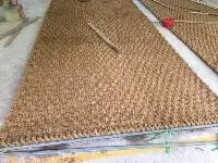
SIZE :
1M X 10 M X 10 MM
1M X 10 M X 14 MM
1M X 10 M X 18 MM
1M X 10 M X 25 MM
1M X 10 M X 32 MM
1M X 10 M X 36 MM
LENGTH: 10 METER
COLOR: BROWN NATURAL
MADE FROM: 100% MEDIUM TOLONG FIBER
IMPURITY: 2% MAX
MOISTURE: 15% MAX
PRICE: NEGOTIABLE
PORT LOADING: HO CHI MINH PORT, VIET NAM
PAYMENT TERM: L/C OR T/T
7899 Surgical X Ray C Arm Plx7200 Suppliers
Short on time? Let Surgical X Ray C Arm Plx7200 sellers contact you.